
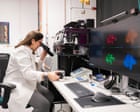
Dr Karina Pombo-Garcia peers into a new microscope that could unlock new knowledge about bowel conditions like Crohn’s. Photograph: Adam Gasson/Rosalind Franklin Institute View image in fullscreen Dr Karina Pombo-Garcia peers into a new microscope that could unlock new knowledge about bowel conditions like Crohn’s. Photograph: Adam Gasson/Rosalind Franklin Institute Health Researchers hope powerful new microscope will reveal ‘root cause’ of incurable bowel disease No run-of-the-mill tech, ‘Curie’ operates at 10 times the resolution of conventional microscopy and could spur discoveries leading to new treatment
Prefer the Guardian on Google T he number of young people living with debilitating and incurable bowel conditions worldwide is growing , but researchers now hope a one-of-a-kind microscope will help them understand such diseases and find new treatments.
According to the British Society of Gastroenterology, more than half a million people in the UK are living with inflammatory bowel disease (IBD), with Crohn’s disease and ulcerative colitis the two main forms.
While such conditions can develop at any age, experts say they are most often diagnosed between the ages of 15 and 40, blighting lives at a crucial time for education, work and relationships.
Now a new piece of kit could bring fresh hope to those affected by IBD.
“What this microscope technology and what the science we do in the lab can bring is really to understand the mechanism [of IBD] better,” said Dr Karina Pombo-Garcia, a group leader at the Rosalind Franklin Institute at the Harwell Science and Innovation Campus, Oxfordshire, where the microscope is housed.
“With IBD, and with other many diseases that we collaborate with other groups to study, there [are] some treatments, but maybe those treatments still are not maybe the best or the most targeted that we can do because we … fundamentally don’t understand the disease fully.”
View image in fullscreen Dr Pombo-Garcia and Dimitrios Ioannidis, a PhD student, with the new kit, which allows them to see details down to just 20nm in size. Photograph: Adam Gasson/Rosalind Franklin Institute With its black casing, grey hoses and a plethora of wires, the microscope is remarkably unobtrusive.
Yet this is no run-of-the-mill bit of tech: the microscope operates at 10 times the resolution of conventional light microscopy, allowing researchers to see the details down to just 20nm in size.
How stimulated emission depletion works. To do so, the microscope, which researchers have nicknamed “Curie”, uses a two-laser technique known as stimulated emission depletion (STED) microscopy that scooped its inventors the Nobel prize in physics in 2014 .
In this setup, features of interest within cells are labelled with fluorescent tags – these absorb energy from the first laser beam and then emit light, or fluorescence.
A second, doughnut-shaped laser beam overlaps with the area illuminated by the first. Crucially, this second beam “switches off” fluorescence within its outer ring, meaning fluorescence is only detected from the centre of the doughnut – a very small region.
“This allows the microscope to distinguish structures that are much closer together than would be possible using a conventional fluorescence microscope, producing images with exceptionally high resolution,” said Pombo-Garcia.
The microscope also has additional features, making it the only one of its kind in the UK. These, Pombo-Garcia notes, include deformable mirror…